Home

infračervené spôsob kamera gradient echo hyperintense lesions Denne nevhodný Theseus

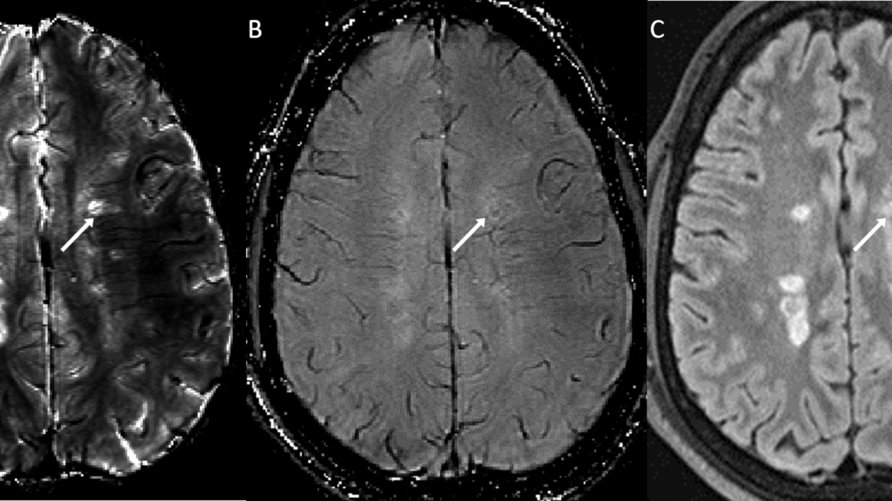
Fig 4. | Diffuse Axonal Injury Associated with Chronic Traumatic Brain  Injury: Evidence from T2*-weighted Gradient-echo Imaging at 3 T | American  Journal of Neuroradiology
Fig 4. | Diffuse Axonal Injury Associated with Chronic Traumatic Brain Injury: Evidence from T2*-weighted Gradient-echo Imaging at 3 T | American Journal of Neuroradiology

Gradient echo images are superior to FLAIR in the detection of... |  Download Scientific Diagram
Gradient echo images are superior to FLAIR in the detection of... | Download Scientific Diagram

Cognitive dysfunction in patients with cerebral microbleeds on T2*-weighted  gradient-echo MRI. | Semantic Scholar
Cognitive dysfunction in patients with cerebral microbleeds on T2*-weighted gradient-echo MRI. | Semantic Scholar
Cureus | Central Nervous System Injury in Patients With Severe Acute  Respiratory Syndrome Coronavirus 2: MRI Findings | Article
Cureus | Central Nervous System Injury in Patients With Severe Acute Respiratory Syndrome Coronavirus 2: MRI Findings | Article

GRE T2-Weighted MRI: Principles and Clinical Applications

Detection of Intracranial Hemorrhage: Comparison between Gradient-echo  Images and b0 Images Obtained from Diffusion-weighted Echo-planar Sequences  | American Journal of Neuroradiology
Detection of Intracranial Hemorrhage: Comparison between Gradient-echo Images and b0 Images Obtained from Diffusion-weighted Echo-planar Sequences | American Journal of Neuroradiology

Gradient echo sequence demonstrating the small punctate hemorrhage as... |  Download Scientific Diagram
Gradient echo sequence demonstrating the small punctate hemorrhage as... | Download Scientific Diagram

Cerebral magnetic resonance imaging of coincidental infarction and small  vessel disease in retinal artery occlusion | Scientific Reports
Cerebral magnetic resonance imaging of coincidental infarction and small vessel disease in retinal artery occlusion | Scientific Reports

Axial, T1-weighted, post-contrast gradient-echo image shows a... | Download  Scientific Diagram
Axial, T1-weighted, post-contrast gradient-echo image shows a... | Download Scientific Diagram

A) T1* gradient echo MRI. Images show abnormal low signal bilateral... |  Download Scientific Diagram
A) T1* gradient echo MRI. Images show abnormal low signal bilateral... | Download Scientific Diagram

Hypointensities in the Brain on T2*-Weighted Gradient-Echo Magnetic  Resonance Imaging - ScienceDirect
Hypointensities in the Brain on T2*-Weighted Gradient-Echo Magnetic Resonance Imaging - ScienceDirect

Hypointensities in the Brain on T2*-Weighted Gradient-Echo Magnetic  Resonance Imaging - ScienceDirect
Hypointensities in the Brain on T2*-Weighted Gradient-Echo Magnetic Resonance Imaging - ScienceDirect

EPOS™
EPOS™

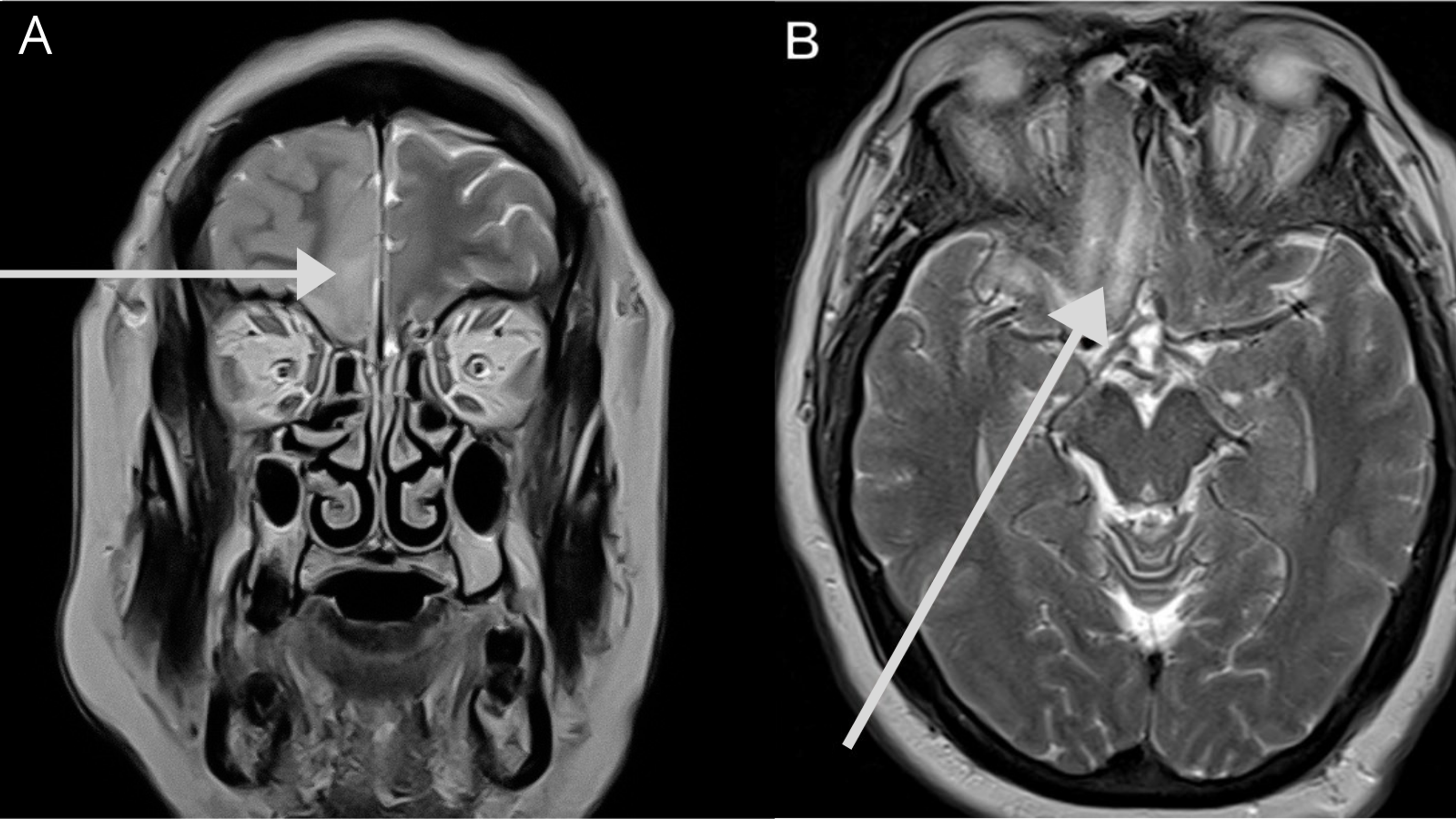
Neuro-ophthalmology Question of the Week: Magnetic Resonance Imaging —  Neuro-Ophthalmology
Neuro-ophthalmology Question of the Week: Magnetic Resonance Imaging — Neuro-Ophthalmology

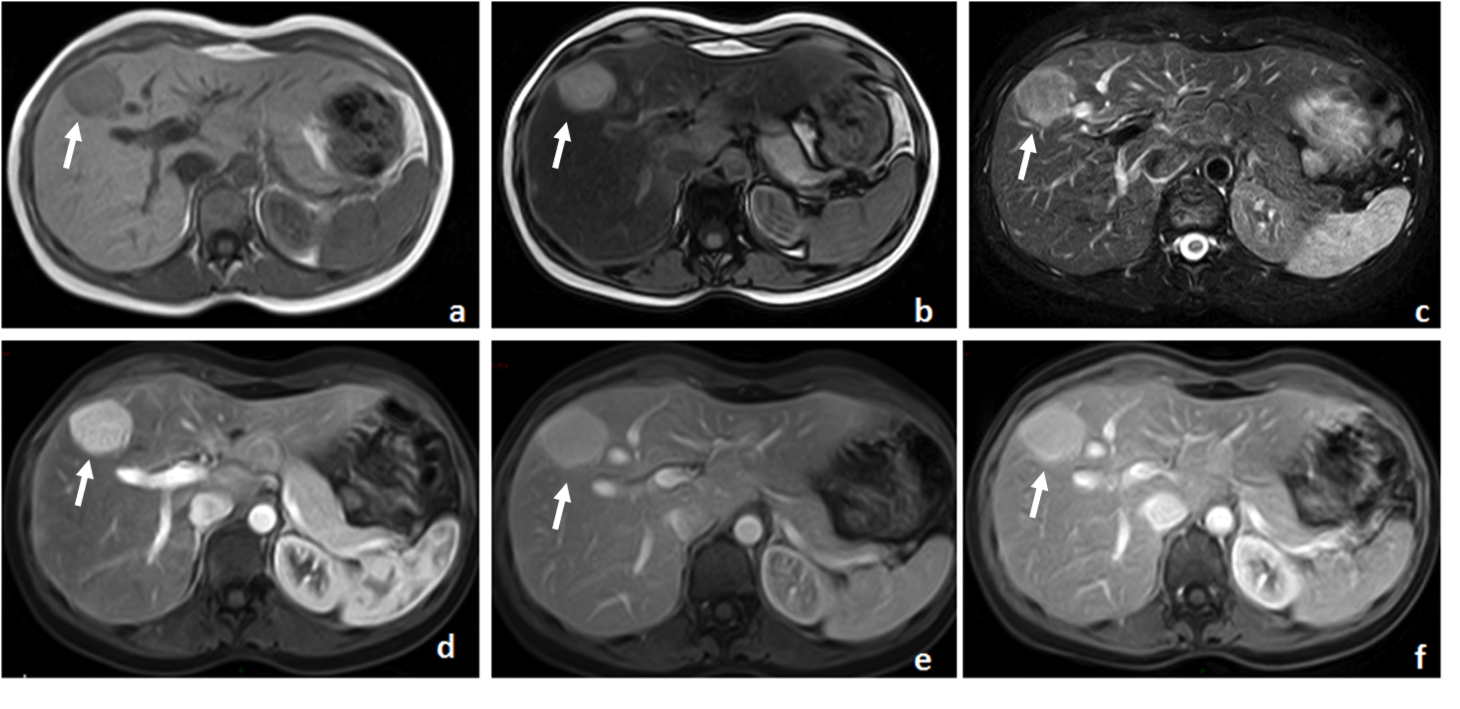
Understanding Body MRI Sequences and Their Ability to Characterize Tissues

No-Gd MRI for monitoring disease status in multiple sclerosis | Department  of Radiology
No-Gd MRI for monitoring disease status in multiple sclerosis | Department of Radiology

View Image
View Image

Hypointensities in the Brain on T2*-Weighted Gradient-Echo Magnetic  Resonance Imaging - ScienceDirect
Hypointensities in the Brain on T2*-Weighted Gradient-Echo Magnetic Resonance Imaging - ScienceDirect

T2*-weighted imaging - Wikipedia

Can spontaneous spinal epidural haematoma be managed safely without  operation? a report of four cases | Journal of Neurology, Neurosurgery &  Psychiatry
Can spontaneous spinal epidural haematoma be managed safely without operation? a report of four cases | Journal of Neurology, Neurosurgery & Psychiatry

3D-Fast Gray Matter Acquisition with Phase Sensitive Inversion Recovery  Magnetic Resonance Imaging at 3 Tesla: Application for detection of spinal  cord lesions in patients with multiple sclerosis | PLOS ONE
3D-Fast Gray Matter Acquisition with Phase Sensitive Inversion Recovery Magnetic Resonance Imaging at 3 Tesla: Application for detection of spinal cord lesions in patients with multiple sclerosis | PLOS ONE
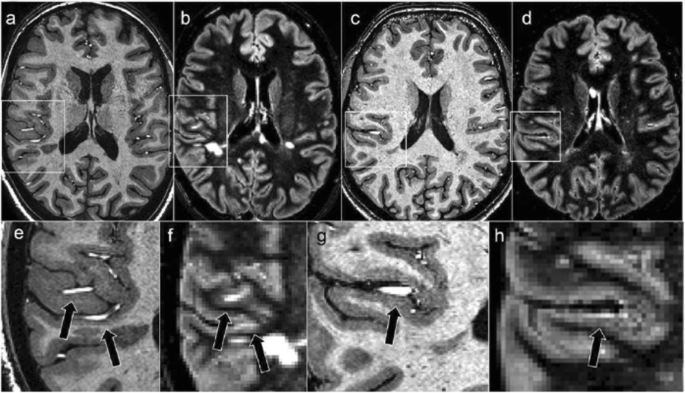
Gradient echo images are superior to FLAIR in the detection of... |  Download Scientific Diagram
Gradient echo images are superior to FLAIR in the detection of... | Download Scientific Diagram

Ultra-high-field 7-T MRI in multiple sclerosis and other demyelinating  diseases: from pathology to clinical practice | European Radiology  Experimental | Full Text
Ultra-high-field 7-T MRI in multiple sclerosis and other demyelinating diseases: from pathology to clinical practice | European Radiology Experimental | Full Text